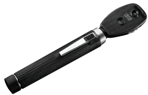
DIRECT OPHTHALMOSCOPE, XHL, 2.5 V
Valid Article
DIRECT OPHTHALMOSCOPE
Definition
A battery-powered, hand-held, ophthalmic instrument designed to examine the interior of the eye allowing the examiner to clearly see the details of the retina and other structures (cornea, aqueous, lens, and vitreous). It consists of a built-in light source that is directed by the examiner through the pupil to illuminate the interior of the eyeball, a mirror with a single view hole, and a dial containing several lenses of different powers that can be alternated by the examiner while viewing. It produces an upright, or unreversed, image of about 15 times magnification.
Specifications
Technical specifications
Direct opthalmoscope
- Handle for batteries
- Screw-on head with all the basic functions
- Different apertures such as: large spot, small spot, half moon, slit, fixation star. The blue interference filter is a must.
- XHL (Xenon halogen) bulb, 2.5 V
- High quality lens
- Supplied with 1 spare bulb
Packaging & Labelling
Delivered in a rigid case with instructions for use.
To be Ordered Separately
Batteries that fit the handle.
must be ordered with a spare-bulb, they are not compatible with other models (with or without blue filter)
Instructions for use
Direct ophthalmoscopes are intended for a transient examination of less than 2 minutes with a 15 minutes break until the next application.
The ophthalmoscope can be adjusted to suit the task at hand. A disc or wheel contains lenses of different powers and the required lens can be brought into the line of sight to correct any refractive error on the part of the patient (or of the user if she is not using her spectacles).
Hold the device as close to the eye as possible
Depending on the instrument, you can choose between different apertures. Minimum available: large spot, small spot, half moon and blue interference filter
- Large/Medium/Small light source: Ophthalmoscopes usually have 2 or 3 sizes of light to use depending on the level of pupil dilation.
- Half light: If, for example, the pupil is partially obstructed by a lens with cataracts, the half circle can be used to pass light through only the clear portion of the pupil to avoid light reflecting back
- Blue light: can be used to observe corneal abrasions and ulcers after fluorescein staining (see related articles)
Precautions for Use
Verify that the lamp voltage complies with the supply voltage of the handle.
Allow the device to cool down before changing the bulb
Storage
- Keep the instrument in its case or pouch when not in use.
- Make sure the on-off switch is fully turned off (a click sound will be heard) before placing the instrument in its case.
- When the ophthalmoscope is not likely to be used for long periods of time, remove the batteries from the handle to avoid leakage.
- While storing the instrument, keep the lens disc on the zero setting so dust does not build up on the other lenses (the zero setting is just a hole with no lens).
- Some ophthalmoscopes include a shutter for the viewing window. It should be closed when the instrument is not in use to prevent dust from entering.
MSF requirements
In practice, this instrument is only used to diagnose serious arterial hypertension (e.g. eclampsia), and very rarely intracranial hypertension.


![[DEXOFLUO1D4] FLUORESCEIN, 0.5%, eye drops, sterile, 0.4ml, unidose amp.](/web/image/product.template/570776/image_256/%5BDEXOFLUO1D4%5D%20FLUORESCEIN%2C%200.5%25%2C%20eye%20drops%2C%20sterile%2C%200.4ml%2C%20unidose%20amp.?unique=c3aebaa)
![[DEXOTROP5D4] TROPICAMIDE, 0.5%, eye drops, sterile, unidose, amp.](/web/image/product.template/570795/image_256/%5BDEXOTROP5D4%5D%20TROPICAMIDE%2C%200.5%25%2C%20eye%20drops%2C%20sterile%2C%20unidose%2C%20amp.?unique=11d80ce)
![[EMEQOPHT1B-] (ophthalmoscope) spare BULB, halogen](/web/image/product.template/572920/image_256/%5BEMEQOPHT1B-%5D%20%28ophthalmoscope%29%20spare%20BULB%2C%20halogen?unique=140966d)